
Characterization of freshwater snail intermediate hosts of schistosomes in four communities from Osun State, Southwest Nigeria

Abstract
Aim: Freshwater snails of the genus Bulinus act as essential intermediate hosts of Schistosoma haematobium, a trematode parasite that causes urogenital schistosomiasis. The snails are widely distributed throughout Nigerian waters. Since species identification of the Bulinus snails is important for appropriate control strategies of urogenital schistosomiasis, this study therefore aimed at identifying the Bulinus species responsible for transmission of the infection in four communities located in an endemic Local Government Area of Nigeria. It also aimed at using restriction fragment length polymorphism (RFLP) as a more affordable method than sequencing to characterize Bulinus snails from schistosomiasis endemic regions in Nigeria.
Methods: In this study, 100 freshwater snails morphologically identified as Bulinus species were collected from four communities located in a previously reported schistosomiasis endemic Local Government Area (LGA), namely Olorunda LGA in Oshun State, Southwest Nigeria. All snails were screened for schistosome infection using polymerase chain reaction (PCR) targeting the DraI gene. Molecular identification of the snails was done by PCR amplification of their entire internal transcribed spacer region including the 5.8S ribosomal RNA gene and RFLP.
Results: Five of the 100 snails were positive for schistosome infection. PCR-RFLP profiles showed bands of different sizes for 26 other snails including the schistosome-infected ones. RFLP analysis showed that 11 of the snails belonged to the freshwater snails of the genus Physa while 13 belonged to the freshwater snails of the genus Bulinus, including Bulinus globosus (8) and B. truncatus (5). The species of the remaining two snails could not be resolved using the reference profiles from our previous studies.
Conclusion: This study confirmed previous observations that limited morphological uniqueness within the Bulinus groups hinders their identification, and RFLP is a cheaper alternative method to sequencing that can be used by laboratories with limited resources for Bulinus species identification.
Keywords
INTRODUCTION
Schistosomiasis is endemic in Africa, the Middle East, and South America[1-5]. In Nigeria, about 29 million people are infected while 101 million people are at risk[6-12]. Bulinus snails are intermediate hosts of Schistosoma haematobium, a trematode parasite that causes urogenital schistosomiasis. The snails are found throughout many parts of Africa and its adjacent regions[13-15]. Identification of Bulinus species based on morphology alone is difficult due to the limited morphological distinctiveness within the species groups.
Several reports have been able to identify and classify species of the snail intermediate hosts into groups based on their physical malacological parameters such as shell appearances and internal structures[16-18]. However, these classifications are sometimes unreliable when used to classify at higher resolution and extremely confusing. Meanwhile, accurate identification of snail-borne trematodiasis is fundamental to the epidemiology and strategic control of the disease, particularly in tropical Africa with the highest burden of schistosomiasis. Consequently, a robust system for identification and classification is required to supplement more traditional approaches.
Enzyme electrophoresis has been used to complement such studies and has proved suitable for identification, clarification of relationships between taxa, and studying aspects of reproductive biology and population[19]. Among the molecular approaches used for the investigation, identification and characterization of Bulinus species includes the examination of variation in ribosomal RNA (rRNA) genes, as determined by conventional restriction fragment length polymorphism (RFLP) analysis and PCR-RFLP of the internal transcribed spacer (ITS)[20]. In addition, the use of randomly amplified polymorphic DNA and sequence analysis of mitochondrial genes has been employed for their identification[21] and infectivity[13-15]. In this study, we aimed to apply PCR-RFLP of the ITS for the identification of Bulinus snail species in four communities located in areas where urogenital schistosomiasis is endemic but such study has neither been carried out nor mass drug administration been performed. We characterized the Bulinus snails by using the RFLP profiles of Bulinus snails from other parts of Nigeria, which had previously been confirmed through sequencing[13] as reference profiles.
METHODS
Study areas
Snail collection was carried out in Ilie, Oree, Oba-Ile, and Oba-Oke communities located within Olorunda LGA of Osun State, Southwest Nigeria between March and June 2019. The study communities were selected based on the previous reports of high prevalence (46%) of urogenital schistosomiasis caused by
Snail samples
In total, 100 snails were collected from Ilie, Oree, Oba-Ile, and Oba-Oke communities between March and June 2019 using a standard scoop net. The snails were washed and preserved in 70% ethanol. Using taxonomic keys[17,18], 100 snails from the four communities [Table 1] were morphologically identified to belong to the genus Bulinus. The snails were then subjected to molecular analysis for schistosome infection status confirmation and species identification at the Molecular Parasitology Research Laboratory of Public Health and Epidemiology Department in the Nigerian Institute of Medical Research, Lagos State, Southwest Nigeria.
Snail sampling sites, their GPS locations, and the number of Bulinus snails examined at each site
| Sampling sites | GPS locations | Number of snails examined |
| Ilie | N-07°58’04.2 E-004°33’04.2 | 36 |
| Oree | N-07°58’01.2 E-004°33’15.3 | 43 |
| Oba-Ile | N-07°58’44.63 E-004°33’18.62 | 16 |
| Oba-Oke | N-07°58’38.838 E-004°33’12.838 | 5 |
| Total | 100 |
Genomic DNA extraction
Genomic DNA (gDNA) was extracted from the tissue of each of the snails using extraction buffer, containing 2-mercaptoethanol, hexadecyltrimethyl-ammonium bromide (CTAB) (solid), tris (hydroxylmethyl) amino-methane, ethylenediaminetetraacetic acid (EDTA), disodium salt solution, and sodium chloride, as follows. Each snail was removed from 70% ethanol and soaked in TE (10mMTris HCL and 1mM EDTA) overnight to remove the remaining ethanol. The tissue was removed from the shell and placed in a sterile 1.5 mL Eppendorf tube, 500 µL of CTAB solution were added and the tissue homogenized. Ten microliters of proteinase K solution were added and incubated at 55 °C for 1 h, with occasional gentle mixing. To each tube was added 500 µL of chloroform/isoamyl alcohol (24:1) to extract genomic DNA. The organic and aqueous layers were mixed gently for 5 min and spun at 20,784 g for 20 min. The upper aqueous layer was removed into another sterile 1.5 mL tube, and an equal volume of 100% ethanol was added, mixed, and incubated at -20 °C overnight. The next day, it was spun at 13,000 rpm for 20 min, and the pellets were washed with 70% ethanol for 20 min. The supernatant was removed, and the pellet was dried at room temperature. When completely dried, it was re-suspended in 50 µL of TE and stored at 4 °C until used.
Molecular screening of snails for schistosome infection
gDNA obtained from each of the snails was subjected to PCR amplification of the schistosome Dra1 repeat using forward primer 5’GATCTCACCTATCAGACGAAAC3’ and reverse primer 5’TCACAACGATACGACCAAC3’ according to previous studies[13,14]. Genomic DNA extracted from adult Schistosoma haematobium obtained from BEI Resources of the National Institute of Allergy and Infectious Diseases (NIAID), National Institutes of Health (NIH), USA was used as positive control, while no template control (NTC) was used as negative control. Each PCR assay was carried out in a total volume of 20 µL containing 5 µL of 5 FIREPol Master Mix (Solis BioDyne, Estonia), 25 pmol of each primer, and 2 µL of gDNA as template. All PCR amplifications were performed with Thermal Cycler (SimpliAmp, Applied Biosystems by Life Technologies, USA) and a thermal profile involving 5 min at 95 °C, followed by 35 cycles of 30 s at 95 °C, 30 s at 59.2 °C, and 30 s at 72 °C, and a final elongation step at 72 °C for 7 min. The amplified products were visualized on 1.5% agarose gel stained with ethidium bromide at a concentration of 0.5 µg/mL. Photo documentation was performed with Gel Documentation and Analysis System (Clinx Science instruments, USA).
Molecular identification of snail species
Molecular identification of the snails was carried out by PCR amplification of their entire ribosomal internal transcribed space (ITS) region including the 5.8S rRNA gene[13]. The forward and reverse primers used were Etts1 5’TGCTTAAGTTCAGCGGGT3’ and Etts2 5’TAACAAGGTTTCCGTAGGTGAA3’. B. globosus gDNA obtained from BEI Resources of NIAID-NIH USA was used as positive control, while NTC was used as negative control. Cycling parameters were 95 °C for 4 min, followed by four cycles of 95 °C for 15 s, 48 °C for 15 s, and 72 °C for 1 min, and then a further 41 cycles of 95 °C for 15 s, 53 °C for 45 s, and 72 °C for 1 min, with the final extension period of 72 °C for 3 min. Amplifications were confirmed through visualization on 1.5% agarose gel using the Gel Documentation and Analysis System (Clinx Science instruments, USA). The PCR products were subjected to restriction fragment length polymorphism (RFLP) by digesting them with RsaI, a 6-base cutting restriction enzyme, in a digestion reaction consisting of 10 µL of PCR product, 1.75 µL of 10 × buffer, 0.75 µL of enzyme, 0.5 µL of bovine serum albumin (BSA), and 2 µL of H2O in a final volume of 15 µL. The digestion mixture was incubated for 30 min at 37 °C. Digestion products were separated by electrophoresis on 1.5% agarose gel stained with ethidium bromide and viewed with the Gel Documentation and Analysis System (Clinx Science Instruments, USA).
RESULTS
PCR amplification of schistosome DraI repeat from 100 snails showed five of the snails to be positive for schistosome infection [Figure 1]. Four of the five infected snails were collected from Ilie and one from Ore. PCR amplification of the entire nuclear ribosomal internal transcribed spacer (ITS) region including the 5.8S rRNA gene of the snails produced different profiles, while PCR products from 26 snails with very good signals on the agarose gels were then subjected to RFLP [Figures 2 and 3]. Bands that were typical of Bulinus species were obtained from 13 of the snails, while 11 produced bands typical of Physa species. However, two infected snails produced different profiles from our previously published reference RFLP profiles for Bulinus snails, which made their species identification unresolved. Using our previously published RFLP profiles for some Bulinus species[13,14] as reference profiles, the RFLP results show that eight of the 13 Bulinus snails were B. globosus and five were B. truncatus [Table 2]. One of the three infected snails from Ilie was identified to belonging to the freshwater snails of the genus Physa and the other two were identified as B. truncatus and B. globosus, respectively. However, the species of one of the four schistosome-infected snails from Ilie and the only infected one from Ore could not be resolved using the RFLP reference profiles from our previous studies.
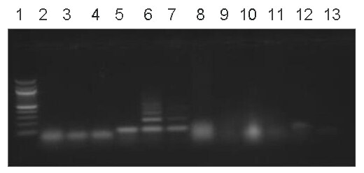
Figure 1. Ethidium bromide-stained agarose gel showing infection status of 10 snails using the schistosoma DraI gene (121 base pair short tandem repeat). Lanes: 1: Quick-Load 100 bp DNA Ladder; 2-4: uninfected snails; 5-7: infected snails; 8 and 11: infection status unresolved; 10: uninfected snail; 12: DNA from adult S. haematobium (positive control); 13: negative control (no DNA).
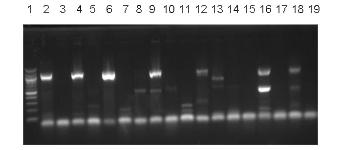
Figure 2. Agarose gel stained with ethidium bromide showing PCR products following amplification of ITS region of 10 snails. Lanes: 1: Quick-Load 100 bp DNA Ladder; 2, 4, 6, and 12: B. truncatus; 8, 10, and 14: Physa; 9 and 16: B. globosus; 13: could not be resolved; 18: B. globosus (positive control); 19: no DNA (negative control). PCR: Polymerase chain reaction; ITS: internal transcribed spacer.
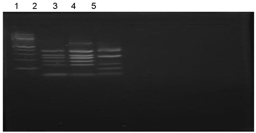
Figure 3. Agarose gel stained with ethidium bromide showing species-specific banding profiles obtained from three of the Bulinus snails through digestion of PCR products of their entire ITS region including the 5.8S rRNA gene. Lanes: 1: Quick-Load 100 bp DNA Ladder; 2 and 3: B. truncatus; 4: B. globosus; 5: negative control. PCR: Polymerase chain reaction; ITS: internal transcribed spacer; rRNA: ribosomal RNA.
Snail species in relation to study locations
| Locations | B. globosus n (infected) | B. truncatus n (infected) | P. acuta n (infected) | Unresolved specie n (infected) | Total infected per site (%) |
| Ilie | 3 (1) | 1 (1) | 5 (1) | 1 (1) | 4 (40%) |
| Oree | 4 (0) | 4 (0) | 3 (0) | 1 (1) | 1 (8.3%) |
| Oba-Ile | 1 (0) | 0 (0) | 2 (0) | 0 (0) | 0 (0%) |
| Oba-Oke | 0 (0) | 0 (0) | 1 (0) | 0 (0) | 0 (0%) |
| Total snails | 8 | 5 | 11 | 2 | 5 |
DISCUSSION
Cercaria sheddings have been used for detection of snail infection with schistosome miracidia, but this method is limited because pre-patent infection can last for several weeks with only a small proportion of the snails making the stage for cercariae shedding[22]. Considering the relationship between the parasite and the snail hosts, evidence-based research focused on accurate identification of the snail host and the parasite would not only define species diversity but also block the transmission, and thus control infection. Therefore, in this study, 100 Bulinus snails were screened for schistosome infection by PCR amplification of the schistosome Dra1. Only five (5%) of the Bulinus snails from Ilie and Ore were infected. This study was carried out in four communities within a schistosomiasis endemic LGA in Southwestern region of Nigeria where such study has not been carried out before. A previous study[13] was done in 28 communities located in three geographical regions of the country, namely North Central, Southeast, and Southwest. This might have accounted for the observation of very few infected snails in the study. The restriction fragment profiles of 13 Bulinus snails - B. globosus (n = 8) and B. truncatus (n = 5) - and 11 Physa species obtained in this study closely matched previously published profiles[10,13,14], as shown by their RFLP profiles [Figure 2]. The Physa genus has been reported[11] to retain schistosome miracidia without shedding or carrying the parasite to patency[13]. This study also confirmed earlier suggestions[13] that restriction fragment analysis of the ribosomal ITS region for species identification of snails in the genus Bulinus could be used as a cheaper alternative to sequencing by laboratories with limited resources. Only 26% of the snails had sufficiently strong PCR products for restriction digest, and 8% of the snails (2/26) could not be taxonomically identified, including one of the infected snails. These limitations were due to the weak PCR products obtained, and the bands were too weak for the RFLP analysis to be carried out. The DNA extraction method used in this study needs to be improved for better PCR products, and further optimization of the PCR may be necessary.
In conclusion, this study confirmed previous observations that limited morphological uniqueness within the Bulinus groups hinders their identification, but RFLP is a cheaper alternative method to sequencing that can be used by laboratories with limited resources for Bulinus species identification[13,14].
DECLARATIONS
Acknowledgment
The authors acknowledge NIH Biodefense and Emerging Infections Research Repository (BEI Resources, USA) for providing the gDNA from B. globosus and adult S. haematobium used as positive controls in this study. The authors are grateful to the Executive Chairman, Olorunda Local Government Area, Osun State, the traditional rulers and the council of elders of Ilie, Ore, Oba-Ile and Oba-Oke for permission to carry out the study in their communities. The cooperation of fishermen and canoe riders of Ilie, Ore, Oba-Ile, and Oba-Oke are also well appreciated. The authors would also like to thank the reviewers for their comments that helped improve the manuscript.
Authors’ contributions
We declare that this work was done by the authors named in this article and all liabilities pertaining to claims relating to the content of this article will be borne by them.
Developed the concept: Akinwale OP, Ugbomoiko US
Designed the experiments: Akinwale OP
Carried out snail collections: Kareem II, Awe DO, Babamale AO
Prepared the samples for molecular assays: Gyang PV, Nwafor TE, Kareem II
Performed molecular assays and data acquisition: Gyang PV, Nwafor TE
Performed data analysis: Akinwale OP
Prepared the manuscript: Akinwale OP, Kareem II, Awe DO, Babamale AO
Takes responsibility for the integrity of this work as a whole from inception to the published article and should be designated as the guarantor: Akinwale OP
Financial support and sponsorship
None.
Conflicts of interest
All authors declared that there are no conflicts of interest.
Ethical approval and consent to participate
Not applicable.
Consent for publication
Not applicable.
Copyright
© The Author(s) 2022.
REFERENCES
1. Health Organization (WHO). Public health impact of schistosomiasis: disease and mortality. WHO expert committee on the control of schistosomiasis. Bull World Health Organ 1993;71:657-62.
2. Stothard JR, Stanton MC, Bustinduy AL, et al. Diagnostics for schistosomiasis in Africa and Arabia: a review of present options in control and future needs for elimination. Parasitology 2014;141:1947-61.
3. Chitsulo L, Engels D, Montresor A, Savioli L. The global status of schistosomiasis and its control. Acta Tropica 2000;77:41-51.
5. Steinmann P, Keiser J, Bos R, Tanner M, Utzinger J. Schistosomiasis, and water resources development: systematic review, meta-analysis, and estimates of people at risk. Lancet Infect Dis 2006;7:411-25.
6. Awogun IA. A comparison of the prevalence and intensity of Schistosoma haematobium and Schistosoma mansoni among secondary schoolchildren in Ilorin, Kwara State. Nigerian J Parasitol 1990;9:51-4.
7. Hotez PJ, Kamath A. Neglected tropical diseases in sub-Saharan Africa: review of their prevalence, distribution, and disease burden. PLoS Negl Trop Dis 2009;3:e412.
8. Hotez PJ, Pecoul B. “Manifesto” for advancing the control and elimination of neglected tropical diseases. PLoS Negl Trop Dis 2010;4:e718.
10. Ude E, Akinwal O, Ukaga C, et al. Prevalence of urinary Schistosomiasis in Umuowele, Agulu community, Anambra state, Nigeria. Int J Health Res 2009;2:4.
11. Ugbomoiko US, Obiezue RN, Ogunniyi TA, Ofoezie IE. Diagnostic accuracy of different urine dipsticks to detect urinary schistosomiasis: a comparative study in five endemic communities in Osun and Ogun States, Nigeria. J Helminthol 2009;83:203-9.
12. Ugbomoiko US, Ofoezie IE, Okoye IC, Heukelbach J. Factors associated with urinary schistosomiasis in two peri-urban communities in south-western Nigeria. Ann Trop Med Parasitol 2010;104:409-19.
13. Akinwale OP, Kane RA, Rollinson D, et al. Molecular approaches to the identification of Bulinus species in south-west Nigeria and observations on natural snail infections with schistosomes. J Helminthol 2011;85:283-93.
14. Akinwale O, Oso O, Salawu O, et al. Molecular characterisation of Bulinus snails - intermediate hosts of schistosomes in Ogun State, South-western Nigeria. Folia Malac 2015;23:137-47.
15. Madsen H, Bloch P, Phiri H, Kristensen TK, Furu P. Bulinus nyassanus is an intermediate host for Schistosoma haematobium in Lake Malawi. Ann Trop Med Parasitol 2001;95:353-60.
16. Owojori OJ, Asaolu SO, Ofoezie IE. Ecology of freshwater snails in Opa reservoir and research farm ponds at Obafemi Awolowo University Ile-Ife, Nigeria. J Appl Sci 2006;6:3004-15.
17. Brown DS, Kristensen TK. A field guide to African freshwater snails II (West African Species). Danish Bilharziasis Laboratory: Chatollenlund Denmark 1993. p. 55.
19. Njiokou F, Bellec C, Jarne P, Finot L, Delay B. Mating system analysis using protein electrophoresis in the self-fertile hermaphrodite species Bulinus truncatus (Gastropoda: Planorbidae). J Moll Stud 1993;59:125-33.
20. Stothard JR, Hughes S, Rollinson D. Variation within the internal transcribed spacer (ITS) of ribosomal DNA genes of intermediate snail hosts within the genus Bulinus (Gastropoda: planorbidae). Acta Trop 1996;61:19-29.
21. Bowles J, Hope M, Tiu WU, Liu X, McManus DP. Nuclear and mitochondrial genetic markers highly conserved between Chinese and Philippine Schistosoma japonicum. Acta Trop 1993;5:217-29.
Cite This Article
Export citation file: BibTeX | RIS
OAE Style
Ugbomoiko US, Kareem II, Awe DO, Babamale AO, Gyang PV, Nwafor TE, Akinwale OP. Characterization of freshwater snail intermediate hosts of schistosomes in four communities from Osun State, Southwest Nigeria. One Health Implement Res 2022;2:88-95. http://dx.doi.org/10.20517/ohir.2022.05
AMA Style
Ugbomoiko US, Kareem II, Awe DO, Babamale AO, Gyang PV, Nwafor TE, Akinwale OP. Characterization of freshwater snail intermediate hosts of schistosomes in four communities from Osun State, Southwest Nigeria. One Health & Implementation Research. 2022; 2(3): 88-95. http://dx.doi.org/10.20517/ohir.2022.05
Chicago/Turabian Style
Ugbomoiko, Uade S., Issa I. Kareem, Doyinsola O. Awe, Abdulkareem O. Babamale, Pam V. Gyang, Timothy E. Nwafor, Olaoluwa P. Akinwale. 2022. "Characterization of freshwater snail intermediate hosts of schistosomes in four communities from Osun State, Southwest Nigeria" One Health & Implementation Research. 2, no.3: 88-95. http://dx.doi.org/10.20517/ohir.2022.05
ACS Style
Ugbomoiko, US.; Kareem II.; Awe DO.; Babamale AO.; Gyang PV.; Nwafor TE.; Akinwale OP. Characterization of freshwater snail intermediate hosts of schistosomes in four communities from Osun State, Southwest Nigeria. One. Health Implement. Res. 2022, 2, 88-95. http://dx.doi.org/10.20517/ohir.2022.05
About This Article
Copyright

Data & Comments
Data


 Cite This Article 10 clicks

 Like This Article 34
likes
Like This Article 34
likes


Comments
Comments must be written in English. Spam, offensive content, impersonation, and private information will not be permitted. If any comment is reported and identified as inappropriate content by OAE staff, the comment will be removed without notice. If you have any queries or need any help, please contact us at support@oaepublish.com.